
Demodex species — cats
Cats can be infested with three species of Demodex: D. cati, D. gatoi, and a third, as yet un-named species.
Overview
Cats can be infested with three species of Demodex: D. cati, D. gatoi, and a third, as yet unnamed species. Demodex cati mites are similar morphologically to D. canis, except that their bodies are narrower. Feline demodecosis associated with D. cati is rare. The localized form of the disease is very similar to that in dogs, most commonly affecting the eyelids, periocular regions and the head and neck, and usually resolves spontaneously. Demodex cati may also cause a ceruminous otitis externa. Generalized demodecosis associated with D. cati is also rare. It is usually associated with underlying disease, for example diabetes, feline leukaemia virus (FLV), or feline immunodeficiency virus (FIV). Lesions are most common on the head, but may also affect the neck, trunk and limbs, and are circumscribed macules and patches of alopecia, scaling, erythema, hyperpigmentation and crusting.
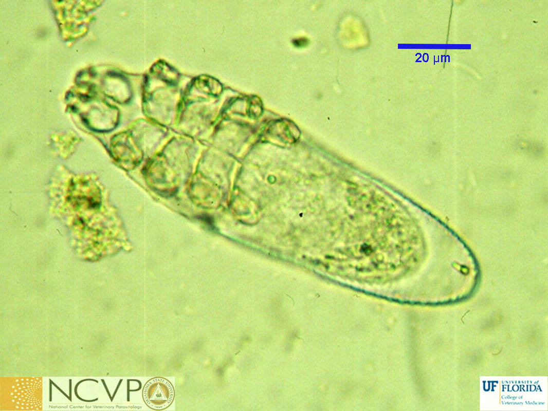
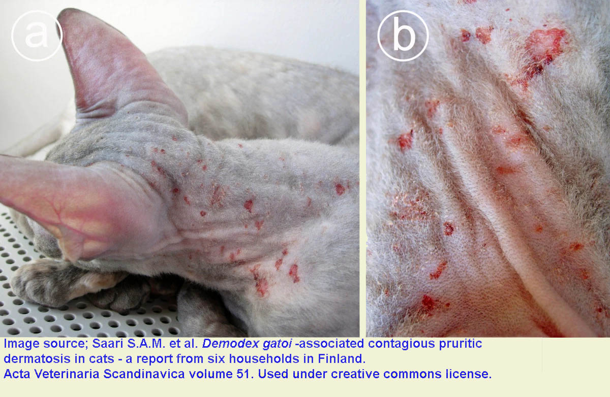
D. gatoi are smaller than D. cati, with a broad, blunt abdomen. The head, neck and elbows are the most commonly affected. The adult mites live in the stratum corneum (more superficially than other Demodex mites) and cause clinical signs suggestive of Notoedres or allergic skin disease, including alopecia, scaling, excoriation, crusting and, unusually, severe pruritus even in the absence of bacterial infection.. Unlike the other species of Demodex, D. gatoi is contagious among cats. The third Demodex species reported from cats resembles D. gatoi but is larger. Little is known of its biology or clinical significance.For diagnosis of demodectic mange in cats, history and clinical signs are very helpful. The diagnosis can be confirmed by detection of mites, and sometimes eggs, in deep skin scrapings. Pinching a skin fold to force the mites from the follicles when the scraping is taken may be helpful. Especially in advanced cases, it may be necessary to examine scrapings from several areas to find the mites. The diagnosis is confirmed if large numbers of adult mites are found, or if there is a large ratio of larvae and nymphs to adults. Demodex gatoi mites are small and translucent and it is easy to miss them on slides prepared from scrapings. Sometimes skin biopsies can help establish a diagnosis of demodecosis, if clinical signs are present but mites cannot be detected in deep skin scrapingCats diagnosed with generalized demodecosis should be tested for FLV and FIV. Treatment is generally not necessary for localized demodectic mange in cats. Isoxazolines effective for canine demodectic mange likely work in cats and should be administered to all in contact cats if D. gatoi is detected. Macrocyclic lactones are inconsistently effective against D. gatoi. Lime sulfur dips and amitraz baths have proven to be effective, if highly inconvenient, treatments for feline demodicosis. All treatments for feline demodecosis are extra label.
People harbour human-specific Demodex mites, particularly in eyelash and eyebrow follicles, without any adverse effects. People cannot be infected with feline Demodex mites
References
Mueller et al. (2020) Diagnosis and treatment of demodicosis in dogs and cats, Clinical consensus guidelines of the World Association for Veterinary Dermatology. Veterinary Dermatology 31:4-e2. https://wavd.org/wp-content/uploads/diagnosis-and-treatment-of-demodicosis-in-dogs-and-cats-mueller-et-al-2020-veterinary-dermatology.pdf
Saari et al. (2009). Demodex gatoi -associated contagious pruritic dermatosis in cats - a report from six households in Finland. Acta Veterinaria Scandinavica 51:40. https://www.ncbi.nlm.nih.gov/pmc/articles/PMC2770525/