
Giardia species
Giardia species occur around the world in people, domestic animal and wildlife.
Summary
Giardia species are flagellated protozoans associated with the intestinal mucosa that occur around the world in people, domestic animals, and wildlife. Dogs can be infected with canine-specific G. canis (Assemblages C and D), as well as with zoonotic G. duodenalis (Assemblage A) and G. enterica (Assemblage B). Infection of dogs with Giardia is much more common than disease, which generally occurs in young animals, often those in sub-optimal environments. Clinical signs usually include diarrhea, dehydration, flatulence and loss of weight.Testing and treatment of dogs for Giardia is generally recommended only for diarrheic animals, or those living in high risk households (immunocompromised people or pets). Diagnosis is based on the detection of trophozoites in direct smears from diarrheic animals,or, more commonly, cysts or cyst antigens in feces.
No drugs are approved in Canada for Giardia spp. in dogs, but metronidazole, fenbendazole, and ronidazole have been used extralabel for the treatment of giardiasis in dogs. Because many dogs are shedding cysts without showing clinical disease and cysts can survive weeks to months in cool, moist environments, re-infection is not uncommon. The goal of treatment is to decrease clinical signs and environmental contamination, not to eliminate infection. Control of Giardia in dogs relies on good sanitation (including bathing affected dogs and disinfecting contaminated indoor environments), minimizing access to sources of infection (surface water, feces of other hosts, including people), and avoiding shared environments with high densities of young dogs. Although there is a potential zoonotic risk, there is scant evidence that dogs are commonly the source of human infections with zoonotic Giardia.
Taxonomy
Phylum Metamonada
Class Trepomonadea
Order Diplomonadida
Family Hexamitidae
Giardia species are mucosoflagellate protozoans but are in a different phylum from other flagellate parasites of importance in animal and human health (e.g., Leishmania, Trypanosoma). Giardia canis (Assemblage C/D) is the host-specific species infecting dogs, and is very rarely zoonotic. Potentially zoonotic species G. duodenalis (certain subtypes of Assemblage A), and G. enterica (Assemblage B) have also been found in dogs.
Note: Our understanding of the taxonomy of parasites is constantly evolving. The taxonomy described in wcvmlearnaboutparasites is based on Deplazes et al. eds. Parasitology in Veterinary Medicine, Wageningen Academic Publishers, 2016.
Morphology
Giardia has two life cycle stages: 1) trophozoites, which live closely attached to the intestinal mucosa; and 2) cysts, which are found in the intestinal lumen and faeces and are the stage by which the parasite is transmitted among hosts.
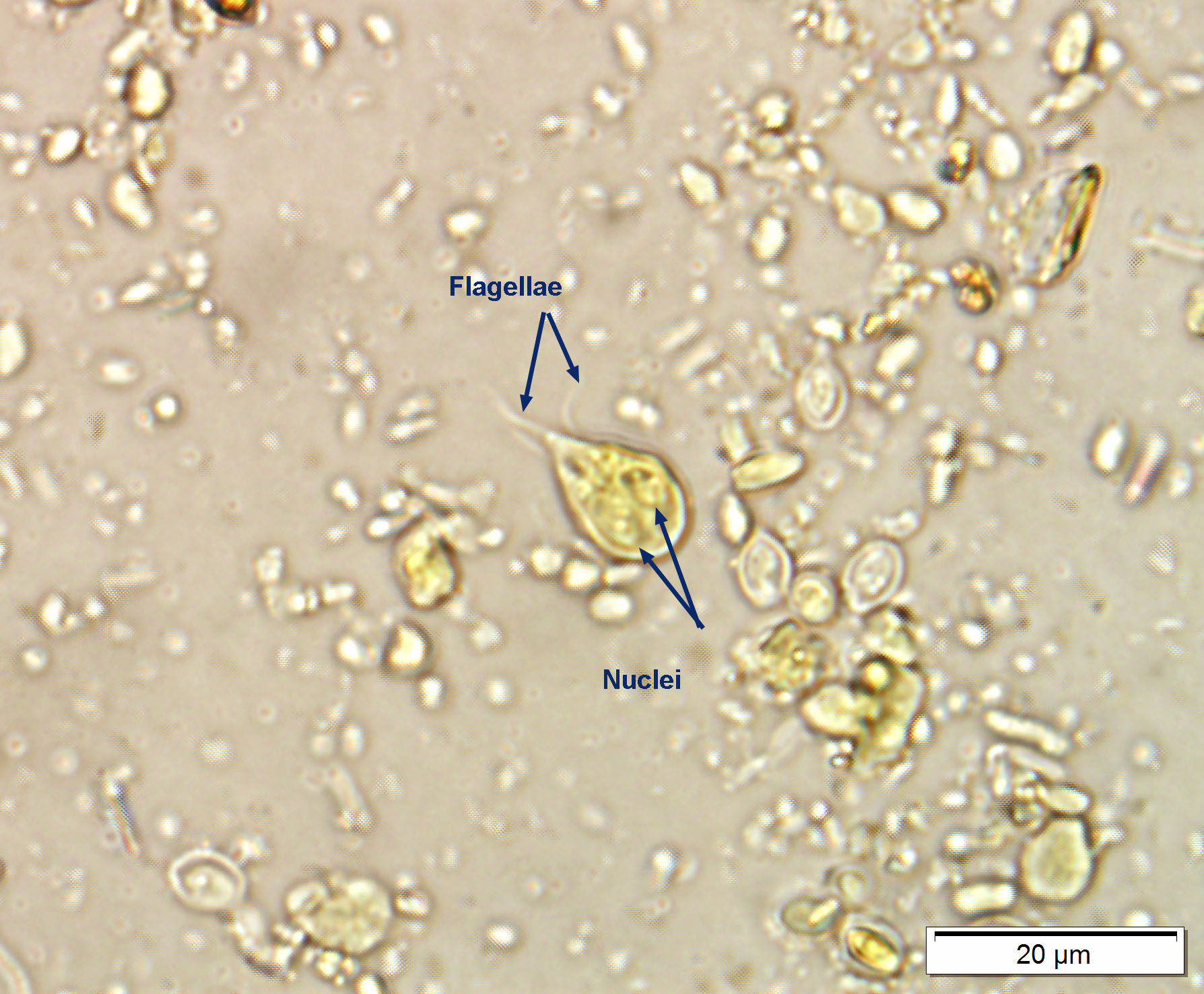
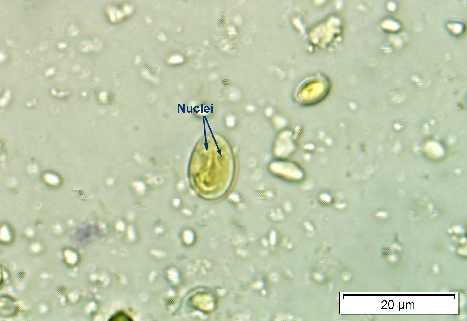
Trophozoites of Giardia are shaped like a pear, and measure approximately 12-15 µm by 5-9 µm. There are two symmetrically arranged nuclei at the broader (anterior) end, one or two comma-shaped median bodies in the mid-section of the trophozoite, four pairs of flagellae (anterior, posterior, caudal and ventral), and a ventral disc which is very important in attaching the trophozoites to the intestinal mucosa. All these structures can be seen by light microscopy, especially in fixed and stained specimens, although usually not all the flagellae are clearly visible. Cysts of Giardia are oval with a smooth cyst wall, and measure approximately 12 µm by 8 µm. Each cyst contains two trophozoites and, therefore, four nuclei grouped at one end of the cyst. Some or all of these nuclei can be seen by light microscopy. There are some morphological differences between the trophozoites and, to a lesser extent, the cysts of the various species, but reliable differentiation requires considerable expertise and experience or molecular techniques.
Host Range and Geographic Distribution
Life Cycle
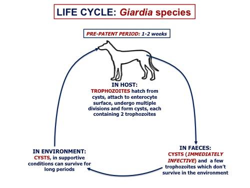
The two stages in the life cycle of Giardia - trophozoites and cysts - have different roles in the life cycle of the parasite. Viable trophozoites are restricted to the lumen of the small intestine, where they are attached to the mucosal surface by means of the ventral disc. Trophozoites are often passed in the faeces of the host, but they die rapidly in the external environment. The stimuli leading the trophozoites to form the cyst stage are not fully understood. Cysts are passed in the faeces of the host and survive very well in the external environment. They are infective when passed by the host, and are the means by which the parasite transmits among hosts.
Giardia trophozoites reproduce asexually by binary fission, which occurs in the intestinal lumen or in newly formed cysts, and each cyst comes to contain two trophozoites. The stimuli leading to cyst formation (encystation) are not fully understood. Once the cysts are ingested by a new host, the cyst wall breaks down and the trophozoites are released into the lumen (excystation). There is evidence from studies of the population genetics of Giardia that the parasite is capable of some form of sexual reproduction, but the details remain obscure. Giardia infections can establish in a host following the ingestion of a very small number of cysts (10). The pre-patent period is usually 1-2 weeks.
Epidemiology
The cyst is the life cycle stage of Giardia that spreads the parasite among its hosts; trophozoites do not survive in the environment. Cysts can occur in very large numbers in host faeces (tens of thousands per gram), especially if the animal has diarrhea (probably indicating a heavy parasite load). Infection and disease are more common in young animals. Cysts can also be present in the hair coat of infected and uninfected hosts. Only a few cysts (10) are required to establish an infection. The cysts of Giardia are very environmentally resistant, surviving in moist, cool conditions for weeks to months, and are relatively resistant to chlorination water treatment . Also, the high proportion of infective hosts that are asymptomatic means they can serve as unrecognized carriers of the parasite. Surveys in several countries show that the overall prevalence of Giardia in dogs is around 10-30%. Environment and host age, as well as test sensitivity and specificity, can affect prevalence data. Immunity is important in clearing Giardia infections, and there is some evidence that infection confers immunity to re-infection.
Transmission is fecal/oral or through consumption of contaminated food and water; water-borne outbreaks are fairly common, especially in summer.
Pathology and Clinical Signs
The life-cycle stage of Giardia responsible for the pathology is the trophozoite, which attaches to the mucosal surface of the enterocytes using the ventral disc. Very rarely, trophozoites might invade the enterocytes themselves or the lamina propria. Giardia often does not cause overt problems, but in some cases, they damage the brush border of the enterocytes, increasing permeability, decreasing absorption and digestion, and altering the intestinal microbiomes. This increased permeability may be why food allergies are occasionally developed in dogs following giardiasis.
Many dogs infected with Giardia show no obvious clinical signs. In most cases, the primary clinical sign is diarrhea (plus or minus mucous or fat), which can range in severity from acute profuse, watery, explosive and foul smelling to more subtle softness. The incubation period is usually one to two weeks, and in the absence of treatment the signs may abate in a couple of weeks. In some animals, the diarrhea tends to be chronic or intermittent. Clinical differences in the effects of the various species of Giardia infecting dogs have not been explored.
Diagnosis
Treatment and Control
It is generally recommended to treat only symptomatic dogs (i.e. diarrheic), or dogs in high risk households (with immunocompromised people or other animals) While several drugs have been shown to be effective in treating Giardia infections on dogs, the most commonly used in Canada are probably fenbendazole (Panacur) and/or metronidazole (Flagyl). Ronidazole is used elsewhere. None are approved for Giardia in dogs in Canada. Following treatment, the parasite might re-appear in the faeces and there may be a recurrence of clinical signs. Both events can be due to recrudescence of parasites that were not removed by the treatment, and/or re-infection from the environment. Therefore, treatment may have to be repeated. The goal of treatment is to decrease clinical signs and environmental contamination, not to eliminate infection, and therefore re-testing following treatment is not generally recommended if clinical signs have resolved. Supportive treatment to deal with the effects of the diarrhea might also be required. Sanitation should include bathing the dog, washing and drying soiled bedding, prompt removal of feces from outdoor areas, and sanitation of soiled indoor surfaces. Cysts are killed by temperatures over 60C, desiccation, UV light, bleach, quarternary ammonium, or hydrogen peroxide solutions. All disinfectants perform best when organic matter is removed and contact times are observed
The control of Giardia in dogs depends on minimizing opportunities for the ingestion of the infective cysts, including preventing dogs from drinking untreated surface water, avoiding shared environments with high density of young dogs, good pooper-scooping practices, and preventing coprophagia. Giardiasis can be a particular problem in groups of dogs kept in sub-optimal conditions, especially pups and animals that are stressed. Because dogs can be infected with zoonotic G. duodenalis and G. enterica, it is very important to advise owners of the zoonotic risk (both ways!). A Giardia vaccine (GiardiaVax®) was approved and marketed for dogs and cats in Canada, but is no longer available.
Public Health Significance
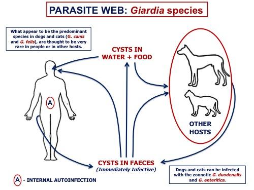
Human giardiasis is commonly called beaver fever, on the (mistaken) assumption that people most commonly acquire the parasite from consuming untreated surface water contaminated with feces from beavers (when instead it is more commonly acquired from human fecal contamination of water). Dogs can be infected with both canine-specific and zoonotic species of Giardia. Commonly used diagnostic tests cannot differentiate species of Giardia, and therefore all canine cases must be treated as potentially zoonotic. In a high risk household (young children or immunocompromised people or pets), it may be worth pursuing species identification, particularly if pet infections with Giardia are refractory to treatment.
Although there is a potential zoonotic risk, there is scant evidence that dogs are the source of human infections with Giardia. More commonly, people acquire Giardia infections from each other (i.e. in daycares or pre-schools), from food, and from potable and recreational water. Outbreaks of human giardiasis from drinking and recreational water are not uncommon in North America, or in other areas of the world.
References
Uehlinger FD et al. (2013) Zoonotic potential of Giardia duodenalis and Cryptosporidium spp. and prevalence of intestinal parasites in young dogs from different populations on Prince Edward Island, Canada. Veterinary Parasitology (In Press)
McDowall RM et al. (2011) Evaluation of the zoonotic potential of Giardia duodenalis in fecal samples from dogs and cats in Ontario. Canadian Veterinary Journal 52: 1329-1333.
Covacin C et al. (2011) Genotypic characterisation of Giardia from domestic dogs in the USA. Veterinary Parasitology 177: 28-32.
Ballweber LR et al. (2010) Giardiasis in dogs and cats: update on epidemiology and public health significance. Trends in Parasitology 26: 180-189.
Payne PA et al. (2009) The biology and control of Giardia spp. and Tritrichomonas foetus. Veterinary Clinics of North America Small Animal Practice 39: 993-1007.
Monis PT et al. (2009) Variation in Giardia: towards a taxonomic revision of the genus. Trends in Parasitology 25: 93-100.
Shukla R et al. (2006) Cryptosporidium spp. and other zoonotic enteric parasites in a sample of domestic dogs and cats in the Niagara region of Ontario. Canadian Veterinary Journal 47, 1179-1184.
Villeneuve, A et al (2015) Parasite prevalence in fecal samples from dogs and cats across the Canadian provinces. Parasites and Vectors 8:281 doi:10.1186/s13071-015-0870-x
https://www.canada.ca/en/public-health/services/laboratory-biosafety-biosecurity/pathogen-safety-data-sheets-risk-assessment/giardia-lamblia.html