
Mites: miscellaneous
A description of a few mites that are found occasionally on dogs and cats.
Nasal mite: Pneumonyssoides caninum
Pneumonyssoides caninum, the nasal mite of dogs and very rarely other carnivores, is a long-legged surface mite that appear pale yellow against the nasal mucosa in life . The adult mites are up to approximately 1 mm in length and 0.5 mm wide and can be seen with the naked eye. Pneumonyssoides caninum is found in dogs around the world, with reported prevalences of up to approximately twenty percent. Individual dogs may harbour up to approximately 250 mites. Many infested dogs show no clinical signs, with some owners being unaware of the mites in their dogs until one or more are sneezed out. Clinical signs of nasal mite infestation include rhinitis-sinusitis, reverse sneezing and watery eyes. There is published evidence that infestation with nasal mites is a significant risk factor for gastric dilatation-torsion. Diagnosis relies on recovery of the mites from sneezed material or by rhinoscopy. Ivermectin, milbemycin, moxidectin and selamectin have been shown to be effective in relieving clinical signs in some cases. These are extralabel uses.
Environmental mites: Dermatophagoides pteronyssimus and D. farinae
These two species of Dermatophagoides are household dust mites, the most common free-living mites associated with skin disease in dogs. Antigens produced by the mites generate a Type 1 hypersensitivity in some dogs, resulting in atopic dermatitis, and the mites are sometimes recovered from the haircoat of atopic dogs. Diagnosis relies on hypersensitivity skin testing, as these mites are ubiquitous. Management as for other allergic skin diseases.
Red mite of poultry: Dermanyssus gallinae
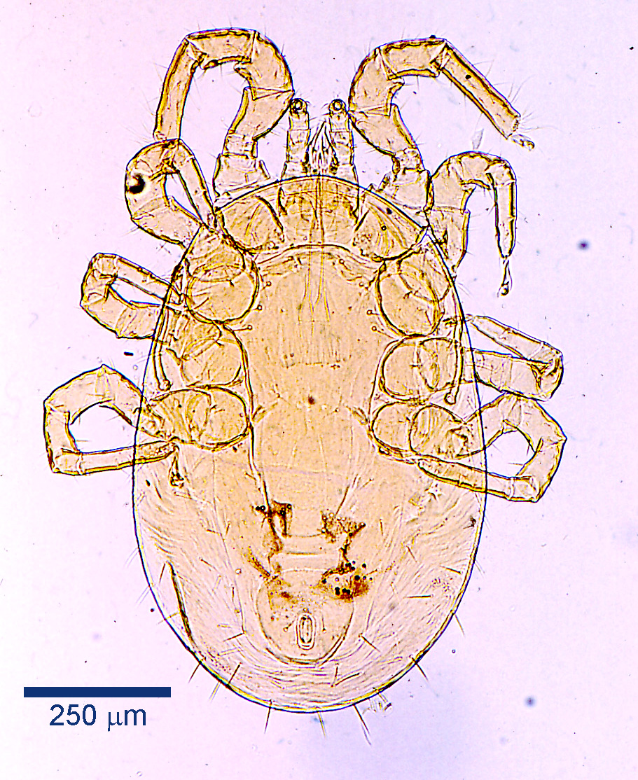
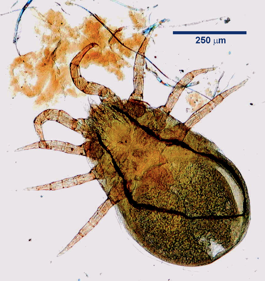
Dermanyssus gallinae, the red mite of poultry – so-called because of its colour when engorged with blood, infests dogs and cats, as well as wild and caged birds and people. The adults are large, measuring up to approximately 1 mm in length. The later nymph stages and the adults of D. gallinae blood feed on their avian and mammalian hosts. Infestation of pets usually results from contact with poultry houses or former poultry houses – the adult mites can survive for many months away from the hosts.
Clinical signs include erythema, papulocrustous eruptions and usually intense pruritus. Diagnosis depends on recovery and identification of the mites. A recent report describes transmission of D. gallinae from cats to people.