
Otodectes cynotis
The ear mite Otodectes cynotis infects dogs and cats and several free-ranging carnivores around the world.
Summary
Taxonomy
Phylum: Arthropoda
Subphylum: Amandibulata
Class: Arachnida
Subclass: Acari
Order: Astigmata
Otodectes species are in the same Order as Notoedres, Sarcoptes, Psoroptes, and Chorioptes (Astigmata), which is different from that containing Cheyletiella, Demodex and Neotrombicula (Prostigmata), and from that containing Dermanyssus and Ornithonyssus (Mesostigmata). Otodectes cynotis is the species in dogs and cats and free-ranging carnivores.
Note: Our understanding of the taxonomy of parasites is constantly evolving. The taxonomy described in wcvmlearnaboutparasites is based on Deplazes et al. eds. Parasitology in Veterinary Medicine, Wageningen Academic Publishers, 2016.
Morphology
Otodectes cynotis is a surface mite and in the adult stage has four pairs of long legs with short pretarsi.
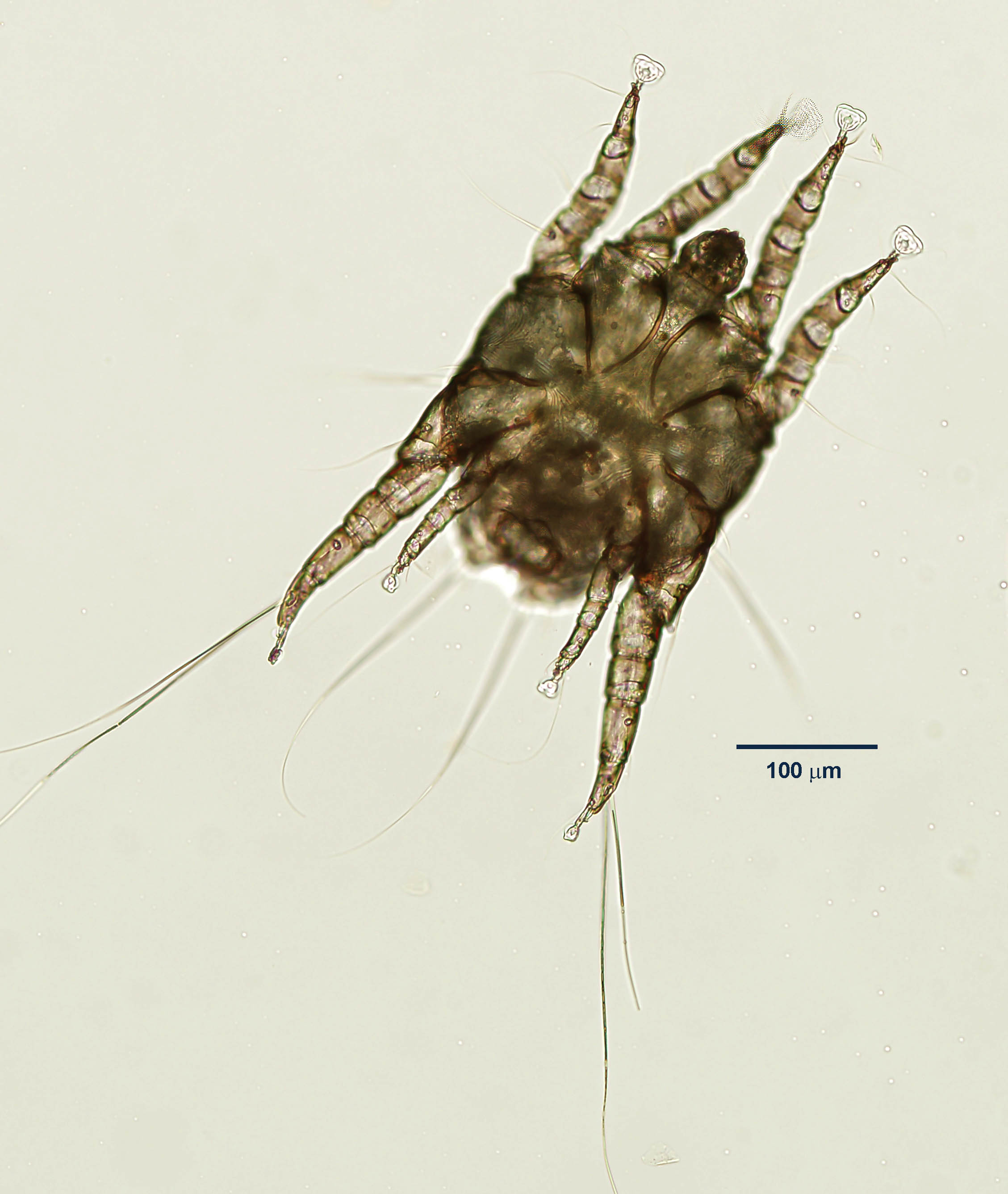
Adult Male
Males have suckers on short pedicels on all legs, females have them only on the first and second pairs of legs.
Adult female Otodectes are 500 x 300 µm in size, and males 395 x 295 µm. Adult mites are barely visible to the naked eye.
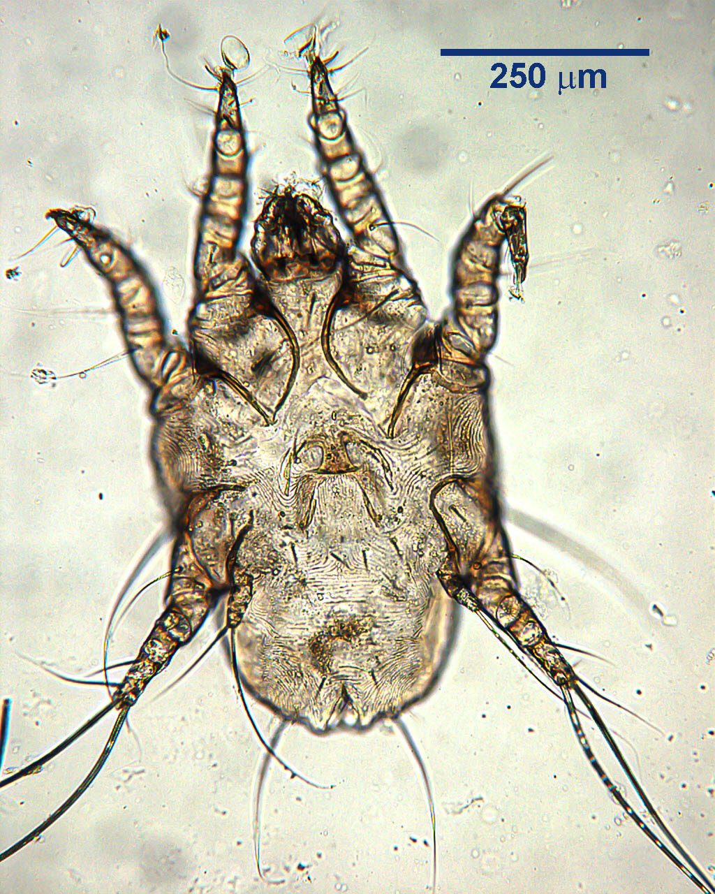
Adult Female
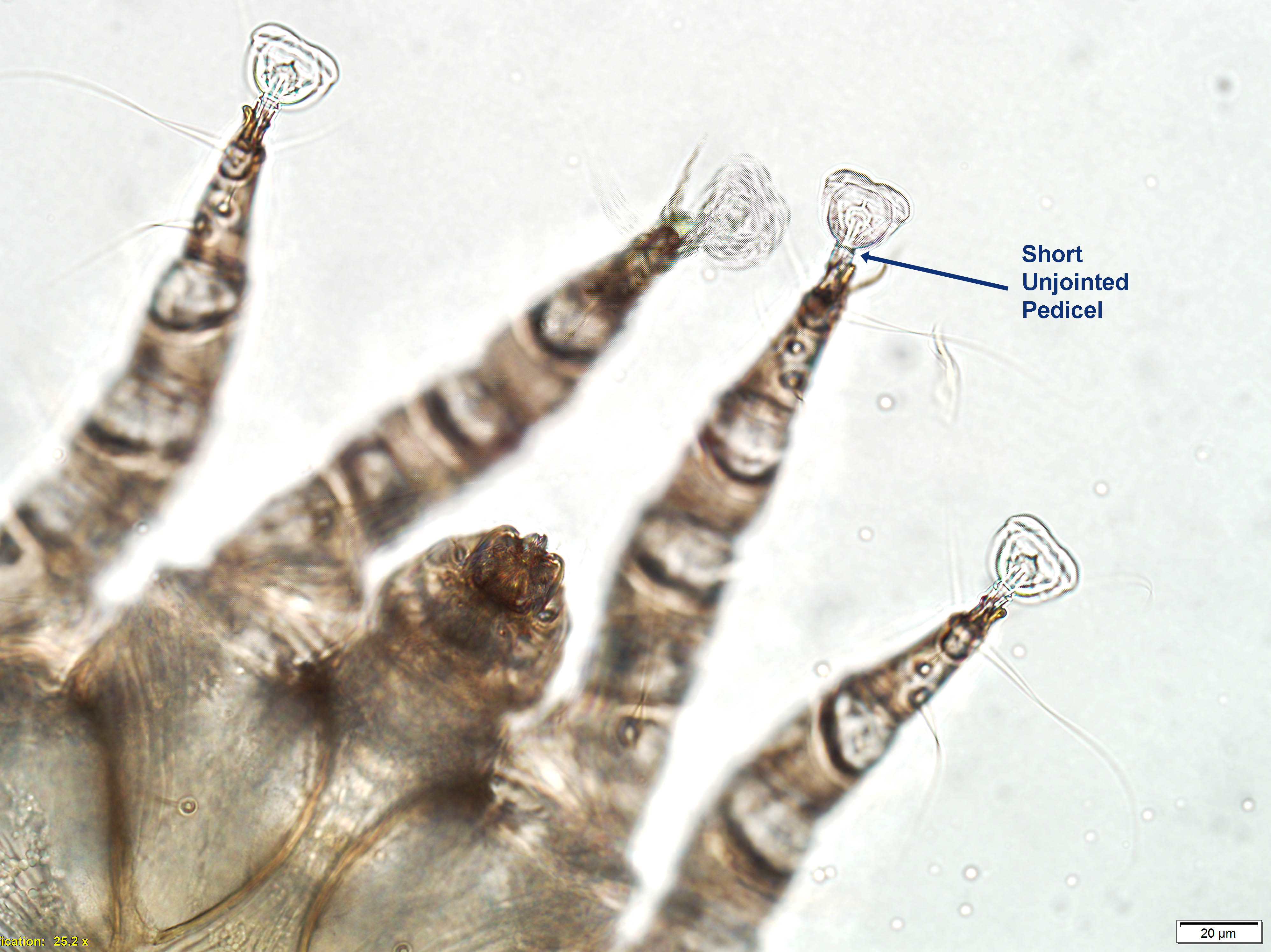
Short Pedicel
Host range and geographic distribution
Otodectes cynotis is found in the ears of dogs and cats, and a variety of free-ranging carnivores, in all parts of the world.
Life cycle
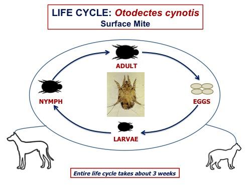
Adult O. cynotis live on the surface of the skin in the external ear canal and the entire life cycle takes place on the host. The adult female mites lay eggs that are cemented to hairs or the skin surface. A six-legged larva hatches from each egg and moults through two eight-legged nymph stages to the adult. The entire life cycle can be completed in approximately three weeks. Adult mites are thought to live for up to approximately eight weeks.
Epidemiology
Otodectes cynotis is very contagious by direct contact or through fomites such as bedding or grooming equipment. Ear mite infestation can spread rapidly in a group of dogs or cats, especially if the hosts are young, immunosuppressed or are in other ways stressed. In most cases, the various life cycle stages may not survive off the hosts for more than a few days under normal environmental conditions, but may survive for months under cool, damp conditions.
Pathology and clinical signs
The mites’ feeding activities and the associated irritation lead to the ear canal filling with skin and mite debris, waxy or crusty – and usually dark brown exudates (“coffee grounds”), and sometimes blood. The lining of the external ear canal is often erythematous. Pruritus is a common feature. Secondary bacterial infection can complicate the mite-induced otitis.
Sometimes mites are found elsewhere than in the ears, for example on the neck, rump and tail, and these mites can be associated with lesions similar to those of flea bite allergic dermatitis.
Dogs and cats may react differently to infestation with O. cynotis, and there may be considerable individual variation. For example, some cats with massive discharge may show no pruritus, and some dogs with few changes in the ears may show severe irritation. Also, some dogs and cats infested with Otodectes may show few or no clinical signs.
Diagnosis
Treatment and control
There are several topical and parenteral products approved in Canada for ear mites in dogs and cats; systemic treatments are better than focal treatment of the ears, as mites can be present elsewhere on the body. Labeled products in Canada include milbemycin (MILBEMITE), moxidectin with imidacloprid (ADVANTAGE MULTI) and selamectin (REVOLUTION). In addition, several products are used extralabel, for example ivermectin and milbemycin (MILBEMAX), and the new isoxazolines, both oral and topical. The mite eggs are not always susceptible to the treatments and, where possible, parenteral treatments with products that persist is preferred. Re-treatment after 3 weeks may be necessary to kill mites that have hatched from eggs in the interventing time period.
Cleaning the ears gently but thoroughly and applying medications to ease the itching, and antibiotics if there is secondary bacterial infection, are also beneficial. It is important to treat all in-contact animals (dogs and cats) because of the rapidity with which ear mites can spread, and to thoroughly clean or destroy bedding and other potential sources of the mites.
Public health significance
References
Robert H. Six, Csilla Becskei, Mark M. Mazaleski, Josephus J. Fourie, Sean P. Mahabir, Melanie R. Myers, Nathalie Slootmans. 2016. Efficacy of sarolaner, a novel oral isoxazoline, against two common mite infestations in dogs: Demodex spp. and Otodectes cynotis. Veterinary Parasitology 222: 62-66, https://doi.org/10.1016/j.vetpar.2016.02.027.
Marília Alves Machado, Diefrey Ribeiro Campos, Natália Lôres Lopes, Isabela Pessôa Barbieri Bastos, Cristiane Bazaga Botelho, Thais Ribeiro Correia, Fábio Barbour Scott, Julio Israel Fernandes. 2018. Efficacy of afoxolaner in the treatment of otodectic mange in naturally infested cats. Veterinary Parasitology 256: 29-31, https://doi.org/10.1016/j.vetpar.2018.04.013.
Adriano F. Vatta, Melanie R. Myers, Jady J. Rugg, Sara Chapin, Aleah Pullins, Vickie L. King, Douglas Rugg. 2019. Efficacy and safety of a combination of selamectin plus sarolaner for the treatment and prevention of flea infestations and the treatment of ear mites in cats presented as veterinary patients in the United States. Veterinary Parasitology 270 (Supplement 1): S3-S11, https://doi.org/10.1016/j.vetpar.2018.11.009.