
Toxascaris leonina
Toxascaris leonina is an ascarid nematode of the small intestine of domestic dogs and cats and free-ranging canids and felids.
Summary
Toxascaris leonina is an ascarid nematode of the small intestine of domestic dogs and cats and free-ranging canids and felids. The parasite occurs around the world, including in Canada, where in dogs in northern regions it to some extent replaces Toxocara canis. Toxascaris leonina has a direct life cycle, although it sometimes uses small mammal paratenic hosts, in which the third stage larva is infective for the definitive hosts. In the environment, the infective stage is the third stage larva in the egg. Following ingestion by a definitive host, these eggs hatch and larvae undergo a mucosal migration prior to completing their development to adults. Diagnosis is based on detecting eggs on fecal flotation and/or adults in feces/vomit, supplemented by coproantigen or coproPCR tests. Treatment of pets is recommended at 2, 4, 6, 8, 12, 16, 20, and 24 weeks of age. Subsequently, treatment for GI nematodes is only recommended for pets at high risk of exposure (free-roaming, coprophagic, raw meat diet, etc), pets in high risk households (young, immunocompromised, or pregnant household members), and pets that test positive on fecal testing. Toxascaris leonina is not known to be zoonotic.
Taxonomy
Phylum: Nematoda
Class: Secernentea
Order: Ascaridida
Superfamily: Ascaridoidea
Family: Ascarididae
Toxascaris leonina is an ascarid nematode, related to the other ascarids in dogs and cats (Toxocara canis and Toxocara cati), and to the ascarids of horses (Parascaris equorum), pigs (Ascaris suum), and people (Ascaris lumbricoides).
The adults of these various ascarids are large to very large, highly fecund, and their eggs are thick-shelled and resistant to adverse environmental conditions. Additionally their life cycles have many similarities, including larval development to the infective stage within the eggs in the environment, and complex larval migration routes in the mammalian hosts.
Note: Our understanding of the taxonomy of parasites is constantly evolving. The taxonomy described in wcvmlearnaboutparasites is based on Deplazes et al. eds. Parasitology in Veterinary Medicine, Wageningen Academic Publishers, 2016.
Morphology
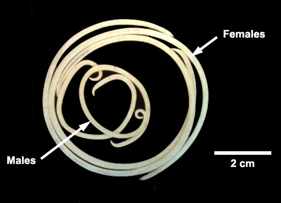
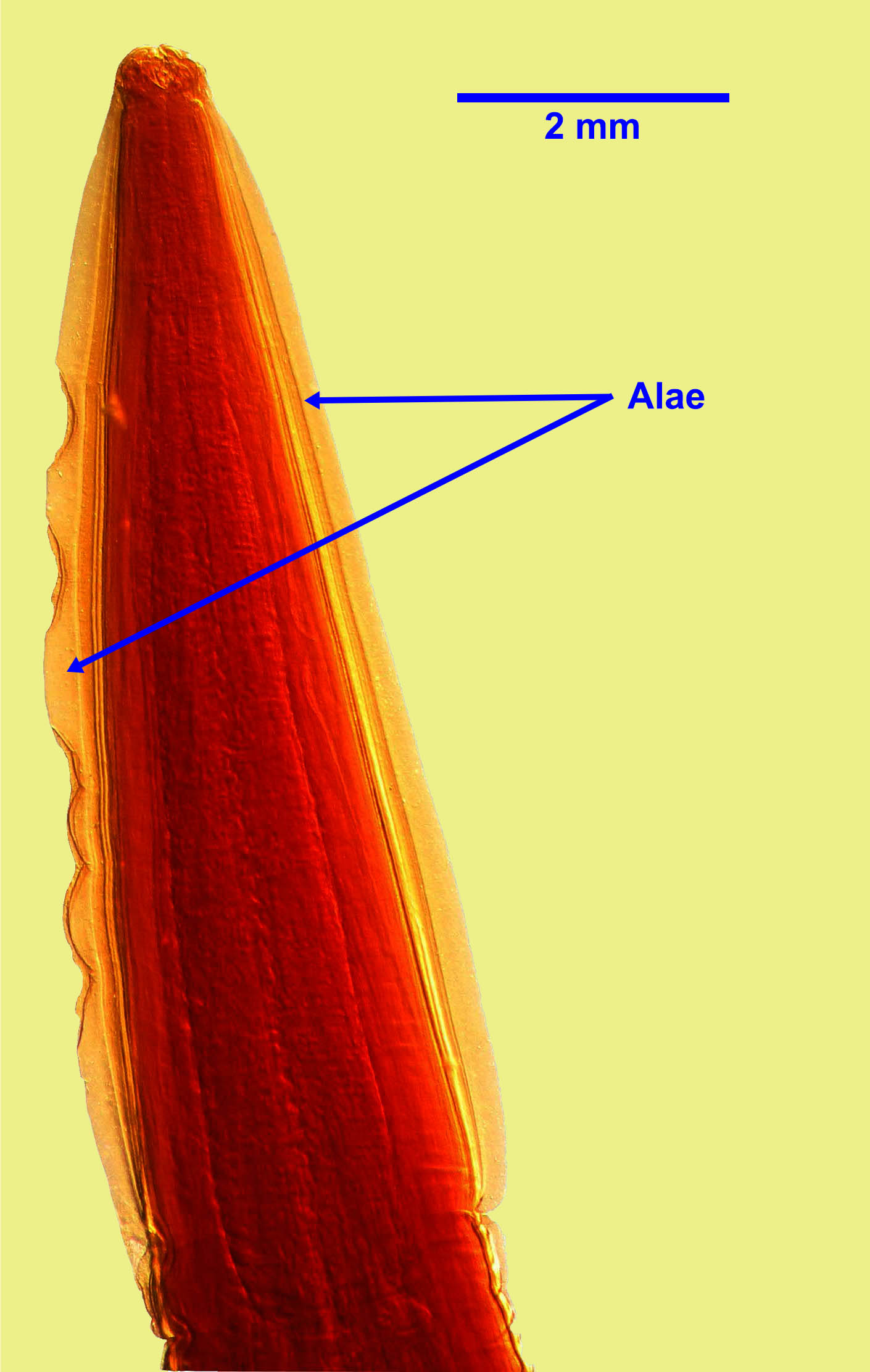
Adult T. leonina are up to approximately 7 cm (males) and 10 cm (females) long and easily visible to the naked eye. Males and females have two long and narrow lateral cervical alae at the anterior end. Males (unlike Toxocara canis) have no finger-like projection at the posterior end.
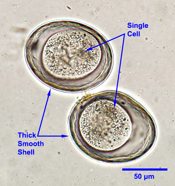
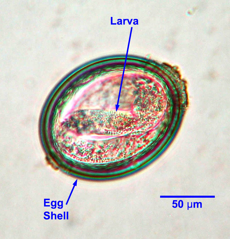
Eggs measure approximately 75 to 85 µm by 60 to 75 µm, and are sub-spherical with a thick, smooth shell (“squash ball” appearance). Each egg contains one or two cells when freshly passed. The eggs can be easily distinguished microscopically from those of T. canis and T. cati.
Host range and geographic distribution
Life cycle - direct
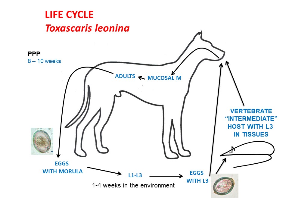
Adult nematodes live in the intestine of the canid or felid definitive host and produce eggs shed in feces. Larvae develop within the eggshell to the infective third stage at a temperature dependent rate (generally 1-4 weeks). Infective third stage larvae within the eggs are ingested by a dog or cat. Then the larvae released undergo a mucosal migration (GI tract lumen to mucosa to lumen) within the GI system (8-10 week prepatent period).
Thus with T. leonina there is neither pre-natal nor trans-mammary transmission, nor somatic larvae. The life cycle may also involve, but does not require, small mammal paratenic hosts, in which third stage larvae hatched from ingested eggs follow a somatic migration (GI tract to portal vessels to liver to heart to lungs to heart to somatic tissues). Third larvae in the tissues of the paratenic host are infective for the dog or cat definitive hosts. Following ingestion by a dog or cat, these larvae too undergo a mucosal migration (8-10 week prepatent period).
Epidemiology
The major route of transmission for T. leonina among dogs is probably through infective eggs and for cats through infected paratenic hosts. Parasite transmission through infective eggs in the environment is facilitated by the properties of the eggs, that are very resistant to adverse environmental conditions and can survive and remain infective for months or perhaps years. Eggs are not immediately infective, but require at least a week in the environment to become infective for definitive and paratenic hosts. Eggs can be inactivated by strong phenol, cresol, or bleach solutions, high temperatures (> 45 C), and desiccation. Eggs are more freeze tolerant than those of Toxocara spp. and freezing at -20 C will not reliably inactivate them. Eggs are not inactivated by most common disinfectants, nor by fixation in formalin or ethanol. The frequent occurrence of T. leonina in dogs in northern Canada suggests that the parasite is well adapted to a relatively cool and harsh environment; it has been detected in Arctic fox well above the Arctic circle.
Pathology and clinical signs
Larval and adult T. leonina have effects in the GI tract similar to those of Toxocara canis and are generally mild unless large number of parasites are present in young animals or in those that are otherwise compromised. In dogs and cats, T. leonina does not migrate beyond the GI mucosa.
Diagnosis
The usually vague clinical signs associated with T. leonina are not helpful for diagnosis. For a specific diagnosis, adult parasites may be found in the feces or vomit, and eggs may be detected on routine fecal flotation, which is relatively sensitive for ascarid eggs. Newer coproantigen and coproPCR methods are increasingly available and useful in detection of ascarid infections, including in the prepatent period. Eggs can often be recovered from adult females. Eggs of T. leonina (smooth shell) are easily distinguishable from those of other nematodes, including Toxocara spp. (rough, pitted shell) and Baylisascaris procyonis (smaller, pitted shell, and more yellow). Adult T. leonina can be distinguished from T. canis and T. cati by the absence of the projection on the tail end of the males and by the eggs in the females, and from T. cati by the shape of the cervical alae.
Treatment and control
Several products are approved in Canada to treat the life cycle stages of Toxascaris leonina in the GI system, and these products are used as part of an overall treatment and control program for GI nematodes. Treatment of pups and kittens for T. leonina is usually less aggressive than for T. canis in pups and T. cati in kittens because of the absence of pre-natal and transmammary infection, and because T. leonina is not zoonotic. Treatment guidelines for Toxocara spp. will be effective against Toxascaris leonina.
Current best practice guidelines from the Canadian Parasitology Expert panel are that pets should be treated at 2, 4, 6, and 8 weeks of age, then monthly to 6 months of age. One of the problems with starting the treatment schedule at two or three weeks of age is that many pups and kittens do not have contact with a veterinarian until they are six or eight weeks old. Providing the drugs to breeders with encouragement towards good compliance would help address this problem.
For pets 6 months and older, recommendations differ depending on the risk profile of the pet and geographic location. In the USA, many parasitologists and veterinarians recommend monthly treatments continuing year-round for the life of the pet. This schedule is thought to increase compliance and also coincide with monthly administration of heartworm preventatives. In most of Canada, where heartworm is not endemic and where infection pressure for other parasites is relatively low, a risk based approach is recommended. Pets which are free-ranging, coprophagic, fed raw meat diets, frequently exposed to heavily contaminated environments, etc are considered high risk for exposure. Owners of service animals and animals in households that contain young children (under 3), pregnant, or immunocompromised people may have higher level of concern about parasitic zoonoses. In these cases (high risk pet and/or high risk household), test 1-2 times annually and treat at least 3-4 times a year. In older, low risk pets in low risk households, test and treat yearly. It is also appropriate to treat only if eggs are detected on fecal examination +/- positive coproantigen or coproPCR tests, which have high sensitivity. Other methods of control include regular removal of pet feces (as infective larvae take at least 1 week to develop), feeding only cooked or commercial diets, and preventing pets from hunting or scavenging wildlife and domestic livestock.
Public health significance
References
Villeneuve, L. Polley, E.J. Jenkins, J.M. Schurer, J. Gilleard, S. Kutz, G. Conboy, D. Benoit, W. Seewald, F. Gagné. 2015. Parasite prevalence in fecal samples from dogs and cats across the Canadian provinces. Parasites and Vectors 8:281 doi:10.1186/s13071-015-0870-x.
Hoopes, J., J.E. Hill, L. Polley, C. Fernando, B. Wagner, J. Schurer, E. Jenkins. 2015. Enteric parasites of free-roaming, owned, and rural cats in prairie regions of Canada. Canadian Veterinary Journal 56(5): 495-501. https://www.ncbi.nlm.nih.gov/pmc/articles/PMC4399737/
Hoopes, J., L. Polley, B. Wagner, E.J. Jenkins. 2013. A retrospective investigation of feline gastrointestinal parasites in western Canada. Canadian Veterinary Journal 54:359-362
Gaunt, M. C., & Carr, A. P. (2011). A survey of intestinal parasites in dogs from Saskatoon, Saskatchewan. Canadian veterinary journal 52(5), 497–500.
Epe C (2009) Intestinal nematodes: biology and control. Veterinary Clinics of North America Small Animal Practice 39: 1091-1107.