
Trictrichomonas foetus
Recognition of the Trictrichomonas foetus as an important enteric parasite of cats first occurred in the mid-1990s. Since then reports from several regions of the world have established that the parasite is an important pathogen in these hosts.
Summary
the mid-1990s. Since then reports from several regions of the world have established that the
parasite is an important pathogen in these hosts. The trophozoites of Tritrichomonas foetus lives
in the ileum and large intestine on the mucosal surface or sometimes in the crypts. They can
cause significant diarrhea. The parasite and disease are seen most commonly in young cats,
especially if overcrowded and/or stressed. Laboratory diagnosis is based on parasite detection in
direct smears of faeces, culture of faeces in selective media, and PCR. Currently there is no
effective treatment. Tritrichomonas foetus has recently been reported from the uterus of a cat in
Norway with uterine pathology. The question of possible transmission of T. foetus between cats
and cattle and vice-versa has not yet been fully resolved.
Taxonomy
Phylum: Parabasalia
Class: Trichomonoda
Order: Trichomonadida
Family: Trichomonadidae
All known members of the Class Trichomonadida are parasitic, and most have only a trophozoite
stage (no cyst) in their life cycles. The taxonomy within the Class is not yet fully established,
particularly at the species level, but molecular approaches are proving helpful.
Tritrichomonas foetus is a flagellate that in cats inhabits the colon. Tritrichomonas foetus in cats
seems to be the same organism as that causing abortion and reproductive losses in cattle, but
more work is required to establish a definitive identification for the species. Organisms from the
two hosts may prove to be genetically and biologically distinct, and there may prove to be several
genotypes, each with their own characteristics, including host specificity. Close relatives of T.
foetus that have a role in human health include Trichomonas vaginalis and Pentatrichomonas
hominis. A less close relative is Giardia.
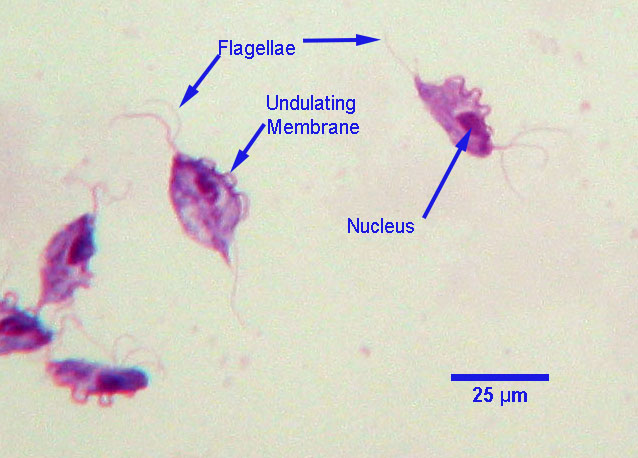
Morphology
and measure up to approximately 25 μm by 15 μm with a nucleus at the broad (anterior) end and
an adjacent sausage-shaped parabasal body. Three anterior flagellae arise from the broad end and
a fourth, posterior, flagellum lies along the outer edge of the undulating membrane before
extending posteriorly. In liquid medium, the trophozoites move with a characteristic tumbling
motion. The flagellae and internal structures can usually be seen in stained and mounted
specimens.
Host range and geographic distribution
Tritrichomonas foetus has been recovered from cattle, pigs and cats in several parts of the world, and may occur in other mammals. The understanding of host range will change as more is learned of the detailed taxonomy of the parasite. A recent study reported a prevalence of 31% among cats at an international cat show in the US.
Life cycle
As far as is known, Tritrichomonas foetus has only a trophozoite stage and transmission is by direct contact, including by ingestion in cats and venereally in cattle. In cats the organism lives in the ileum, caecum and colon, mostly close to the mucosal surface but also occasionally in the mucosal crypts.
Epidemiology
Little is known of the epidemiology of Tritrichomonas foetus in cats. Young, densely housed cats seem to be most commonly infected and affected clinically. It is assumed there are carrier cats that act as a source of infection for others, and transmission from mothers to kittens may be important for maintenance of the parasite in cat populations. The significance of possible host-switching (for example from cats to cattle and vice versa) is not known.
Pathology and clinical signs
Trophozoites of T. foetus damage the mucosa of the large intestine by mechanisms that have not yet been determined. Typical lesions include lymphocytic-plasmacytic colitis, epithelial cell hypertrophy in the crypts, and disruptions of the surface epithelium. Where these lesions are particularly severe, organisms can invade the lamina propria and deeper layers of the intestinal wall. Tritrichomonas foetus has recently been reported from the uterus of a cat in Norway with uterine pathology.
The characteristic clinical sign of feline tritrichomoniasis large bowel diarrhea. Cats in shelters and purebred show cats are more often infected than house cats, but infection is believed to be more common than is clinical disease. Because there are currently no effective treatments for T. foetus, the infection in cats often becomes chronic, often lasting months before the parasite disappears spontaneously.
Diagnosis
History and clinical signs are often helpful. In addition, three laboratory techniques are used to detect T. foetus in feces: a) direct smear and visualization of the trophozoites (the least sensitive); b) fecal culture in the In Pouch™ TF Feline; and c) PCR (the most sensitive). The infection has not yet been recognized with sufficient frequency in western Canada for any of these techniques to have been widely applied.
Treatment and control
There are no drugs approved in Canada for the treatment for Tritrichomonas foetus in cats, and none of those tried (extralabel) has been found to be effective.
Because so little is known about the parasite’s epidemiology, it is difficult to recommend effective control measures. The high prevalences reported in shelters and among purebred show cats suggest that avoidance of overcrowding, good environmental hygiene and minimizing stress may be helpful.
Public health significance
Tritrichomonas foetus is not known to be zoonotic, although there is a single case report of infection with an organism described as T. foetus in the CSF and urine of a person who had been given an allogenic peripheral blood stem cell transplant. According to the authors of the report, however, it is possible that the parasite in this case may have been T. mobilensis or another trichomonad.
References
Payne PA et al. (2009) The biology and control of Giardia and Tritrichomonas foetus. Veterinary Clinics of North America Small Animal Practice 39: 993-1007.
Stockdale HD et al. (2006) Feline trichomoniasis: an emerging disease? Compendium on Continuing Education for the Practicing Veterinarian 28: 463-471.
Foster DM et al. (2004) Outcome of cats with Tritrichomonas foetus infection. Journal of the American Veterinary Medical Association 225: 888-892.