
Hitachi SU8010 Field-Emission Scanning Electron Microscope (FE-SEM)
About
The Hitachi SU8010 is a semi-in-lens type cold field emission FE-SEM. It offers ultra-high resolution imaging of sample surface, and energy dispersive spectroscopy and backscattered electron detection for compositional information.
This SEM is also capable of correlative light-electron microscopy, enabling researchers to investigate details of their samples with SEM in relation to images/information collected using light microscopy.
Features and specifications
- Magnification at high magnification mode: 100x-800,000x
- Magnification at low magnification mode: 20x-2,000x
- Accelerating voltage: 0.5-30kV
- Landing voltage in deceleration mode: 0.1-2.0kV
- Imaging of surface structure
- Detection of backscattered electrons for z-atomic contrast
- Energy Dispersive Spectroscopy using the AztecLiveStandard with Ultim Max 170 Detector
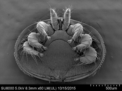
Image examples
Image: Drs. Sarah Wood and Elemir Simko, WCVM.
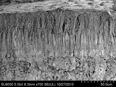
Image: Drs. Marina Leis and Bruce Grahn, WCVM.
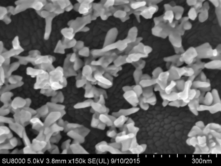
Image: Sajna Simon and Dr. Ian Burgess, USask Dept. of Chemistry.
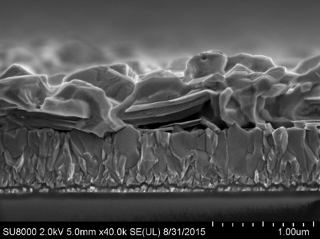
Image: Kianoosh Poorkazem and Dr. Timothy Kelly, USask Dept. of Chemistry.
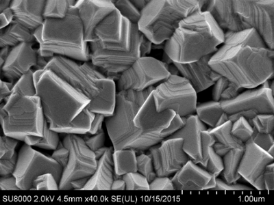
Image: Kianoosh Poorkazem and Dr. Timothy Kelly, USask Dept. of Chemistry.