
Confocal laser scanning microscopes
Zeiss 710, equipped with a two-photon imaging system
Features and specifications
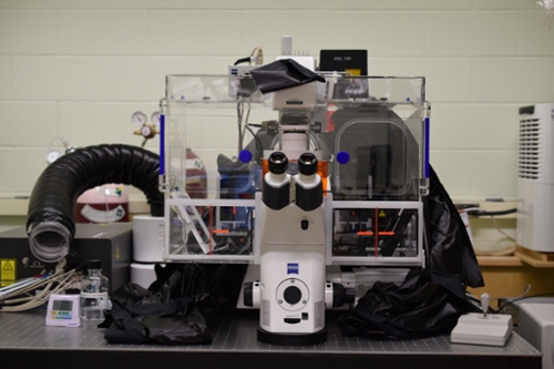
- Equipped with a Coherent Chameleon two-photon system with a tuning range of 705-980 nm, suitable for deep tissue imaging (out of service until further notice)
- A selection of excitation lasers for conventional confocal imaging (458, 488, 514, 543, and 633 nm excitation lasers)
- Spectral imaging and linier unmixing of overlapping signals
- A humidified environmental chamber with five per cent CO2 supply and temperature control for live cell imaging
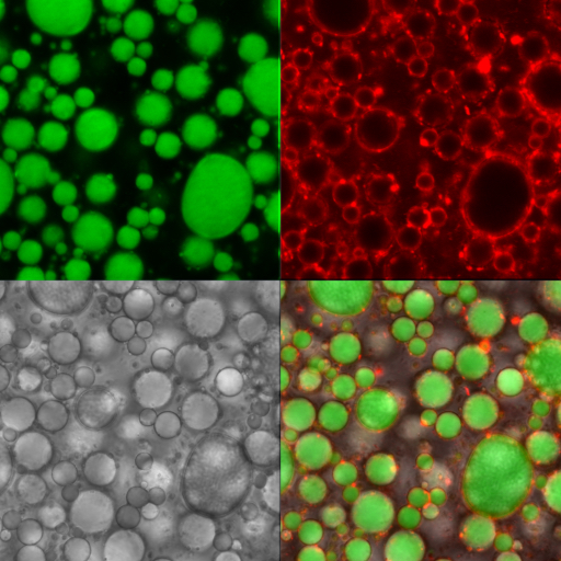
Image examples
Courtesy of Cathy Coutu, Dr. Thushan Withana-Gamage and Dr. Janitha Wanasundara (AAFC).
Fungus (red) penetrating root epidermis (green), three-dimensional rendering with Imaris.
Courtesy of Cathy Coutu and Dr. Gary Peng (AAFC). PLOSONE (2014) vol9, issue 4: e94144.