
Pelodera (Rhabditis) strongyloides
All life cycle stages of the nematode Pelodera strongyloides are free-living. Adults and larvae can, however, invade the skin of dogs and other mammals.
Summary
Taxonomy
Class: Secernentea
Order: Rhabditida
Family: Rhabditidae
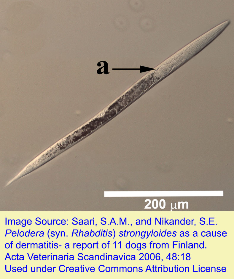
Pelodera strongyloides (previously Rhabditis strongyloides) is a free-living nematode that is in the same order as Strongyloides spp. Larvae and occasionally adults can invade the skin of a variety of mammals..
Note: Our understanding of the taxonomy of helminth, arthropod, and particularly protozoan parasites is constantly evolving. The taxonomy described in wcvmlearnaboutparasites is based on Deplazes et al. eds. Parasitology in Veterinary Medicine, Wageningen Academic Publishers, 2016.
Morphology
Host range and geographic distribution
Pelodera strongyloides has been recovered from the skin of dogs, cattle, sheep, people, and probably other animal hosts, in several areas of the world. Several cases in dogs have been observed in Canada.
Life cycle - free living
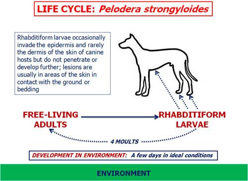
Pelodera strongyloides is a free-living nematode that is an occasional parasite. Adult males and females live in the environment and produce rhabditiform larvae that invade the epidermis and rarely the dermis of the skin, but do not penetrate or develop further. Normally, the rhabditiform larvae in the environment undergo four moults as they develop to the free-living adult stage.
Epidemiology
Pelodera strongyloides is a free-living nematode; thus, the environment is the sole source of infection. The parasite most probably does not transmit from one animal to another. Environmental conditions (i.e. wet, decomposing plant matter) that are supportive of the survival and development of the parasite probably enhance the risk of infection in animals.
Pathology and clinical signs
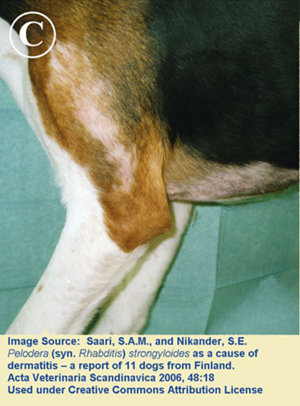
The pathology and clinical signs associated with P. strongyloides result from host reaction to the parasites. Skin lesions are usually localized to areas in contact with the ground. There is usually erythema, alopecia and variable pruritus, and there may also be scaling, crusting and signs of secondary bacterial infection.
Diagnosis
The history and clinical signs are often very helpful. Pelodera strongyloides thrives in less than clean environments that provide nutrition for the adult parasites and conditions that enhance survival of the free-living stages. Thus it is possible that one or more individuals in a group of animals sharing some element(s) of an environment may show similar clinical signs.
A definitive diagnosis of P. strongyloides is usually based on the recovery of typical rhabditiform larvae, and sometimes also adult parasites, from deep skin scrapings. A skin biopsy may also be helpful, but only if parasite stages are fortuitously present in the sections. Other potential etiologies for larvae associated with ventral dermatitis in dogs include the hookworms Ancylostoma caninum and Uncinaria stenocephala; Pelodera larvae are generally larger than hookworm larvae. Pelodera larvae from skin scrapings that are placed in a Petri dish containing suitable nutrient agar will quickly develop to adults and reproduce successfully. Seeking the parasites in the environment is usually unrewarding and it is difficult to distinguish among the various species of free-living nematodes that may be present.
Treatment and control
The first step in treatment is to remove the affected animal(s) from the source of infection, which should then be thoroughly cleaned or, if possible, destroyed. Bathing the animal(s) in a soothing shampoo will probably help the healing process. Treatments with anti-parasitic products are usually not indicated, but ivermectin has helped in some published cases. Removal of the animals from the source of the nematode commonly results in rapid resolution of the clinical signs.
Public health significance
Pelodera strongyloides has been reported from people with skin lesions in several areas of the world. As with animals, the source of infection is the environment. It has been suggested that people can acquire P. strongyloides from affected animals, especially dogs with which they have close contact, but this seems unlikely.
References
Saari SAM et al. (2006) Pelodera (syn. Rhabditis) strongyloides as a cause of dermatitis: a report of 11 cases from Finland. Acta Veterinaria Scandinavica 48: 48.18.
Clark EG, Griffin S, Goodall P. Saskatchewan. Pelodera dermatitis in a dog. Can Vet J. 1989 Dec;30(12):970. PMID: 17423483; PMCID: PMC1681327.