
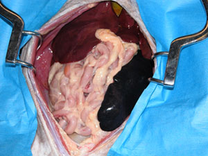
Systematic abdominal exploration
Goal: to thoroughly, but efficiently, examine all of the structures in the abdomen.
- Balfour self-retaining retractors are helpful to hold abdomen open
- exploration should be done first (assuming patient is stable)
- ensure it gets done
- surgical decisions made with all the facts
- specific order that structures are examined varies with surgeon
- do same way each time so it becomes automatic
- avoids forgetting something
- do same way each time so it becomes automatic
Once the entire abdomen has been explored, biopsies or other samples are collected as indicated and the definitive surgical treatment is performed.
- when no abnormalities are found, biopsies should generally include:
- liver, stomach, duodenum, jejunum, ileum, lymph node and bile for culture
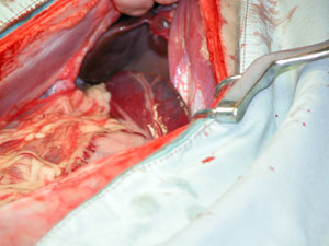
Diaphragm
- thorough examination requires removal of the falciform fat
- gently retract liver caudally
- examine the diaphragm
- specific order that structures are examined varies with surgeon
- do same way each time so it becomes automatic
- avoids forgetting something
- do same way each time so it becomes automatic
Anatomy Review
Some examples of abnormalities that might be identified include:
- metastatic cancer
- diaphragmatic hernia (above)
Anatomy check: Can you identify the structures labelled in the diagram?
Diaphragm Test
Liver
- palpate and visually inspect each lobe
- note changes in size, texture and edges
- express gallbladder and check patency – can usually see dilation of the bile duct and its opening on the duodenum
Anatomy check: Can you identify the structures labelled in this diagram?
Anatomy Review
Some examples of abnormalities that might be identified include:
- liver masses (neoplasia, cirrhosis, abscess)
- inflammation or necrosis of the gallbladder
- choleliths
- bile duct obstruction (stones, neoplasia)
- multiple or single portosystemic shunts
- lymph node enlargement or discoloration
Anatomy check: Can you identify the structures labelled in this diagram?
Liver Test (PDF)
Liver Answer (PDF)
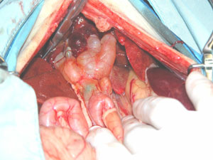
GI tract and associated structures
- examine both visually and by palpation
Anatomy Review
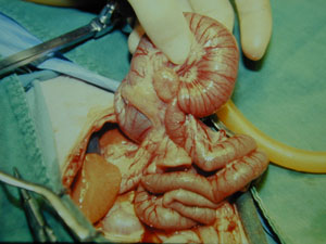
- intestine
- feel as well as look when running the intestines (Movie)
- also examine the associated mesentery, vessels, lymphatics and lymph nodes at same time
- duodenocolic ligament
- limits ability to exteriorize duodenum
- right lobe of pancreas within mesoduodenum
- when reach duodenocolic ligament
- gently flip intestines over to right and follow duodenum to the jejunum, ileum, cecum and colon
OR
- feel as well as look when running the intestines (Movie)
- move directly to the ileocecocolic junction and examine colon and then return to cecum and move orally from
- ileum to jejunum to remaining duodenum until reach the ligament ileum recognized by its antimesenteric vessels and the ileocecal fold
- click here (Movie) to see how to replace them when finished running intestine
Examples of some common abnormalities include:
- foreign bodies or intussusception
- neoplasia
- ulceration
- lymph node enlargement (reactive, neoplastic)
- inflammation, thickening or adhesions
- abnormal sized or positioned organs (volvulus, intussusception, ileus)
- distended lymphatics
- abnormal vascular supply
Anatomy check: do you know the anatomy of this region?
Right trough (paravertebral)
- mesoduodenum* is retracted toward left side of the abdomen so the right paravertebral region can be examined
- NOTE: some surgeons will examine these structures while evaluating the duodenum
Anatomy Review
Some examples of abnormalities that could be detected in this region
- pancreatitis or pancreatic neoplasia
- metastatic masses or defects in the abdominal wall
- abnormal portosystemic communications
- adrenal or renal enlargement or masses ureteral distention or stones
- ovarian follicles or corpus luteum
- pregnancy, pyometra* or masses
Anatomy check: Can you identify the structures labelled in this diagram?
Right Trough Test (PDF)
Right Trough Answer (PDF)
Left trough (paravertebral) and pancreas
Mesocolon (mesentery associated with the descending colon) is used to retract abdominal organs towards right side of abdomen so the left trough or paravertebral region can be examined:
- NOTE: some surgeons will examine some of these structures as they are examining the stomach and the colon
-
Anatomy Review
Some examples of abnormalities that could be detected in this region:
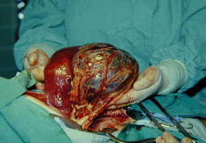
- splenic tumor (hemangiosarcoma)
- metastatic neoplasia or adhesions on the greater omentum
- splenic masses or malpositioning (torsion)
- pancreatitis or masses
- metastatic masses or defects in the abdominal wall
- adrenal or renal enlargement or masses
- dilation of the ureter or stones
- ovarian follicles or corpus luteum
- pregnancy, pyometra* or masses
Anatomy check: Can you identify the structures labelled in this diagram?
Left Trough Test
Pelvic region
Structures to examine include:
- bladder and distal ureters
- distal colon
- iliac lymph nodes prostate and vas deferens or uterine body, cervix and cranial vagina
- urethra (generally not easily evaluated)
- inguinal rings with associated vessels and nerves and vas deferens in males
-
Anatomy Review
Some examples of abnormalities that could be detected in this region include:
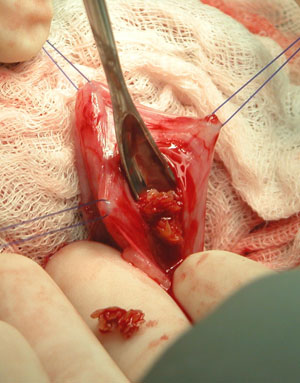
bladder stones being removed during a cystotomy
- bladder thickening, inflammation, stones and masses
- colonic masses
- lymph node enlargement or masses
- prostatic enlargement, masses or inflammation
- cryptorchid testicles
Anatomy check: Can you identify the structures labelled in this diagram?